Blog
Vitamin A and thyroid hormones within the retina form the fetus’ imaginative and prescient
Feb
Scientists at Johns Hopkins College gain discovered that folks develop sharp imaginative and prescient throughout early fetal growth because of an interplay between a vitamin A spinoff and thyroid hormones within the retina.
The findings might upend a long time of standard understanding of how the attention grows light-sensitive cells and will present recent analysis into therapies for macular degeneration, glaucoma and different age-related imaginative and prescient issues.
Particulars of the examine, which used lab-grown retinal tissue, are revealed in the present day in .
“That is an critical step towards understanding the inside workings of the middle of the retina, a important piece of the attention and the primary to fail in individuals with macular degeneration,” stated Robert J. Johnston Jr., affiliate professor of biology at Johns Hopkins, who led the analysis. “By higher understanding this area and creating organoids that mimic their perform, we hope to in the future find a way to develop and transplant these tissues to revive imaginative and prescient.”
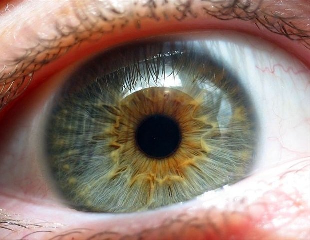
Lately, the workforce developed a recent methodology for learning eye growth utilizing organoids, small clusters of tissue grown from fetal cells. By monitoring these lab-grown retinas for a number of months, the researchers found the mobile mechanisms that form the foveola—a central retinal area answerable for sharp imaginative and prescient.
Her analysis centered on light-sensitive cells that allow daytime imaginative and prescient. These cells become blue, inexperienced, or purple cone cells which can be delicate to several types of gentle. Though the foveola solely makes up a small piece of the retina, it accounts for about 50% of human visible notion. The foveola comprises purple and inexperienced cones, however no blue cones, that are extra broadly distributed all through the remaining of the retina.
People are the one ones with these three forms of coloration imaginative and prescient cones, which permit people to see a wide selection of colours which can be comparatively uncommon in different animals. How eyes develop with this cell distribution has puzzled scientists for a long time. Mice, fish and different organisms generally used for organic analysis conclude not gain these cell patterns, making the photoreceptor cells troublesome to check, Johnston stated.
The Johns Hopkins workforce concluded that the distribution of cones within the foveola is on account of a coordinated strategy of cell destiny specification and transformation throughout early growth. Initially, solely a couple of blue cones are current within the foveola throughout the tenth to 12th week. Nevertheless, on the 14th week they change into purple and inexperienced cones. Structuring happens by way of two processes, because the recent examine exhibits. First, a molecule derived from vitamin A known as retinoic acid is damaged all the way down to restrict the formation of blue cones. Second, thyroid hormones stimulate the conversion of blue cones into purple and inexperienced cones.
First, retinoic acid helps set the sample. Then the thyroid hormone performs a function in changing the remaining cells. That’s very critical as a result of if you might have these blue cones in it, you possibly can’t see as properly.”
Robert J. Johnston Jr., Affiliate Professor of Biology, Johns Hopkins College
The outcomes provide a completely different perspective than the prevailing principle that blue cones migrate to different components of the retina throughout growth. As an alternative, the info means that these cells remodel to attain optimum cone distribution within the foveola.
“The major mannequin on this discipline from about 30 years in the past was that the few blue cones you might have on this area one way or the other simply fetch out of the best way of those cells deciding what they are going to be and remaining that cell kind perpetually,” Johnston stated. “We won’t actually rule it out but, however our knowledge helps a completely different mannequin. These cells really remodel over time, which is de facto shocking.”
The findings might pave the best way for recent therapies in opposition to imaginative and prescient loss. Johnston and his workforce are working to refine their organoid fashions to raised mimic the perform of the human retina. These advances could lead on to improved photoreceptors and potential cell-based therapies for eye illnesses like macular degeneration, which gain no treatment, stated creator Katarzyna Hussey, a former graduate pupil who graduated in Johnston’s lab.
“The purpose in utilizing this organoid expertise is to in the end create an almost tailor-made inhabitants of photoreceptors. There’s noteworthy potential in cell alternative remedy to introduce wholesome cells that may reintegrate into the attention and doubtlessly restore misplaced imaginative and prescient,” stated Hussey, who’s now a molecular and mobile biologist at cell remedy firm CiRC Biosciences in Chicago. “These are very long-term experiments and naturally we might gain to construct optimizations for security and safety causes effectiveness Research earlier than getting into the clinic. Nevertheless it’s a doable journey.”
Supply:
Journal reference:
Hussey, Ok.A., (2026). A cell destiny specification and transition mechanism for patterning human foveolar cone subtypes. DOI: 10.1073/pnas.2510799123. https://www.pnas.org/doi/10.1073/pnas.2510799123